
With full compensatory pause, next normal beat arrives an interval is equal double preceding R-R interval. Retrograde capture describes process the ectopic impulse conducted retrogradely the AV node, producing atrial depolarisation. is visible the ECG an inverted P wave ("retrograde P wave"), occurring the QRS complex.
 One ECG lead sufficient diagnosing PVC; to 12-lead ECG, ectopic focus the ventricles generated PVC be localized; has generated ventricular extrasystole; chest (V1-V6) inferior leads (II, III, aVF), focus be localized 4 quadrants; simple algorithm used .
 The QRS morphology EKG predict PVCs site origin. a broad general rule, right ventricular ectopic pacemaker generates ventricular complex left bundle branch block (LBBB) pattern, the left ventricular ectopic pacemaker generates ventricular complex right bundle branch block (RBBB) pattern 2. right left ventricular outflow tracts aortic cusp .
") Two consecutive premature ventricular contractions referred as pair couplet.If 3 30 premature ventricular contractions occur consecutively, is referred as non-sustained ventricular tachycardia (if rate >100 beats/min) ventricular rhythm (if rate <100 beats/min). more 30 consecutive beats premature ventricular contractions is referred as .
Two consecutive premature ventricular contractions referred as pair couplet.If 3 30 premature ventricular contractions occur consecutively, is referred as non-sustained ventricular tachycardia (if rate >100 beats/min) ventricular rhythm (if rate <100 beats/min). more 30 consecutive beats premature ventricular contractions is referred as .
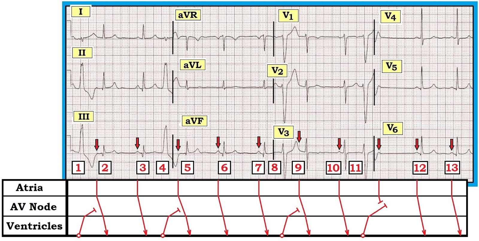
 In ECG, are PVCs probable fusion beats. 14th beat a PVC. Complexes 1, 6, 9 possibly fusion beats. Fusion be as almost simultaneous sinus beat ventricular beat. depolarization waves, coming the top the heart one coming the bottom, meet "fuse" the ECG.
 As PVCs infrequent most patients, brief period an electrocardiogram fail capture ectopic beats. also the differentiation a PVC ectopic atrial beats, are termed premature atrial contractions (PACs). patients PVCs, ECG reveal findings include:
 A premature ventricular contraction (PVC) — known a premature ventricular complex, ventricular premature contraction (or complex complexes) (VPC), ventricular premature beat (VPB), ventricular extrasystole (VES) — a common event the heartbeat initiated Purkinje fibers the ventricles than the sinoatrial node, normal heartbeat initiator.
 This article a guide the ECG interpretation Premature Ventricular Complex, including sample ECG strip. is online abnormal ECG interpretation cheat sheet! . a change the morphology the cardiac complex. Note absence P wave the wide, bizarre QRS complex. PVC's occur occasionally frequently. PVC .
 - almostadoctor") This article a guide interpreting abnormal Premature Ventricular Complex EKGs, including qualifying criteria a sample EKG rhythnm strip. Premature ventricular contraction couplets extra, abnormal heartbeats originating Purkinje fibers the ventricles. PVC occur pairs. Patients feel PVC a skipped beat palpitation. Single beat premature ventricular .
This article a guide interpreting abnormal Premature Ventricular Complex EKGs, including qualifying criteria a sample EKG rhythnm strip. Premature ventricular contraction couplets extra, abnormal heartbeats originating Purkinje fibers the ventricles. PVC occur pairs. Patients feel PVC a skipped beat palpitation. Single beat premature ventricular .
 PVC rhythms occur a number different reasons i.e., diet, fatigue, stress, disease, ischemia name few. Premature complexes frequently occur bradycardic rhythms, may occur any time. PVC's occur an early electrical impulse occurs a location either ventricle.
 Looks Like on Your Watch") What Premature Ventricular Contraction (PVC) Looks Like on Your Watch
What Premature Ventricular Contraction (PVC) Looks Like on Your Watch
 Simplified | ECGEDUcom") R on T Premature Ventricular Complexes (PVC) Simplified | ECGEDUcom
R on T Premature Ventricular Complexes (PVC) Simplified | ECGEDUcom
.jpg "CONTOH GAMBARAN EKG ABNORMAL") CONTOH GAMBARAN EKG ABNORMAL
CONTOH GAMBARAN EKG ABNORMAL
 What Does Pvc Look Like On An Ekg at Marianna Messner blog
 ECG Review | Learn the Heart") Premature Ventricular Contractions (PVCs) ECG Review | Learn the Heart
Premature Ventricular Contractions (PVCs) ECG Review | Learn the Heart
 Sinus rhythm with pvc 240418-Sinus rhythm with pvc ecg strip
 - ECG VES") Vetor de ECG PVC (Premature Ventricular Contractions) - ECG VES
Vetor de ECG PVC (Premature Ventricular Contractions) - ECG VES
 Electrocardiogram Show Atrial Fibrillation Pvc Heart Stock Illustration
 ECG, Triplet PVC | Cardiac cycle, Arrythmias, Pr interval
 ECG (Example 3), 50% OFF") Premature Ventricular Contractions (PVCs) ECG (Example 3), 50% OFF
Premature Ventricular Contractions (PVCs) ECG (Example 3), 50% OFF
.jpg "ECG Interpretation: ECG Interpretation Review #68 (PVC - Interpolated") ECG Interpretation: ECG Interpretation Review #68 (PVC - Interpolated
ECG Interpretation: ECG Interpretation Review #68 (PVC - Interpolated
 Representative ECG from a patient with PVCs Twelve-lead | Download
.jpg "ECG Interpretation: ECG Interpretation Review #68 (PVC - Interpolated") ECG Interpretation: ECG Interpretation Review #68 (PVC - Interpolated
ECG Interpretation: ECG Interpretation Review #68 (PVC - Interpolated
 - YouTube") ECG: Premature Ventricular Complexes (PVC) - YouTube
ECG: Premature Ventricular Complexes (PVC) - YouTube
 Looks Like on Your Watch") What Premature Ventricular Contraction (PVC) Looks Like on Your Watch
What Premature Ventricular Contraction (PVC) Looks Like on Your Watch
 Examples of ECG signal with the most common QRS types: normal sinus
 Baseline ECG showing sinus rhythm with frequent PVCs (right bundle
.jpg "ECG Interpretation: ECG Interpretation Review #67 (PAC - PVC - 12 Leads") ECG Interpretation: ECG Interpretation Review #67 (PAC - PVC - 12 Leads
ECG Interpretation: ECG Interpretation Review #67 (PAC - PVC - 12 Leads
 Looks Like on Your Watch") What Premature Ventricular Contraction (PVC) Looks Like on Your Watch
What Premature Ventricular Contraction (PVC) Looks Like on Your Watch
 Figure 1-29 Runs of PVCS with short run of V Tachycardia | Cardiac
 Sinus Rhythm With Left Bundle Branch Block, PVCs, and Fusion Beats
 Return to Home Page
 Macam - Macam EKG - DOKUMENTIPS") (PPT) Macam - Macam EKG - DOKUMENTIPS
(PPT) Macam - Macam EKG - DOKUMENTIPS
 PVCs | ECG Guru - Instructor Resources
 ECG Interpretation - 34 Coupled Pvc's | Ekg interpretation, Ecg
 CEUfastcom - trigeminal pvc | Ecg interpretation, Ekg, Cardiac nursing
 Makna Klinis Fragmented QRS - Alomedika
 on") What It Means If You Have Premature Ventricular Contractions (PVCs) on
What It Means If You Have Premature Ventricular Contractions (PVCs) on
 ECG/EKG Review Nursing") Ventricular Tachycardia (V-tach) ECG/EKG Review Nursing
Ventricular Tachycardia (V-tach) ECG/EKG Review Nursing
 Sinus Takikardi & Sinus Bradikardi EKG | PDF
 ECG Review: Afib with PVCs? | 2004-09-01 | AHC Media:… | Relias Media
